Eye Surgery Complications and Coverage Options
Helpful resources
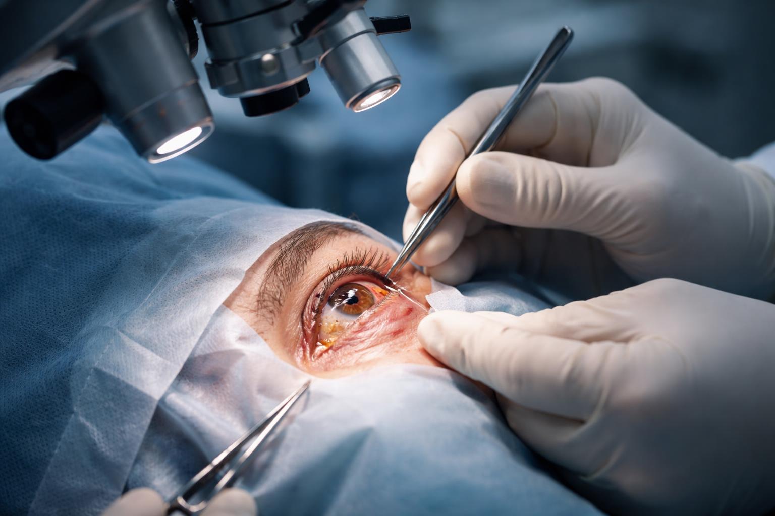
Eye Surgery Complications Explained: Risks, Prevention, and Recovery
Eye surgery, while generally safe and effective, carries risks that patients should understand before proceeding. Common complications include infection, inflammation, and vision disturbances, which can occur during or after the procedure. Serious complications are rare, but knowing what to watch for helps ensure timely care and better outcomes.
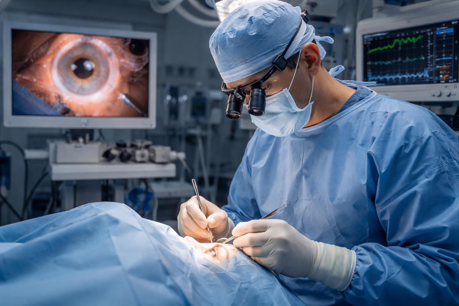
The types of risks vary depending on the surgery performed, such as cataract removal or refractive procedures like LASIK. Surgeons, including ophthalmologists and optometrists, take precautions to minimize these risks, but patients must also follow post-operative instructions carefully. Understanding potential issues promotes informed decisions and realistic expectations.
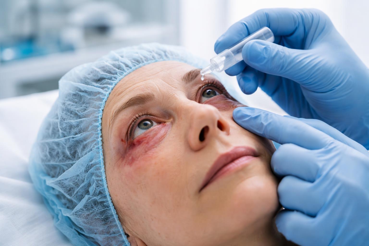
Close monitoring by eye care professionals is crucial after surgery to identify and address any problems early. Some complications can be managed effectively, preventing long-term damage to vision and eye health.
Key Takeways
- Complications can occur but serious problems are uncommon.
- Different surgeries carry distinct risks and side effects.
- Professional follow-up and patient care reduce adverse outcomes.
Causes and Risk Factors
Complications after eye surgery arise from multiple factors tied to patient health, surgical methods, and individual ocular history. Understanding these elements helps in identifying patients at higher risk and tailoring preventive strategies.
Underlying Health Conditions
Chronic diseases such as diabetes and hypertension significantly increase the risk of postoperative complications. Diabetes, for instance, can impair wound healing and raise the likelihood of infection or inflammation after surgery.
Hypertension affects vascular health and may contribute to unexpected bleeding or poor ocular perfusion during and after procedures. Patients with systemic illnesses often require thorough preoperative assessment by an ophthalmologist to manage these risks effectively.
Additionally, ocular surface diseases, including dry eye and tear film abnormalities, compromise the eye’s natural defense mechanisms. Such conditions increase irritation, delay healing, and lead to inflammation, making surgery more complex and raising the chance of adverse outcomes.
Type of Surgery Performed
Different eye surgeries carry distinct risk profiles. Cataract surgery, while generally safe, can be affected by flap tears or corneal scars, particularly during retreatment or complicated procedures. The use of faulty instruments or improper technique can worsen the risk of flap-related complications.
Laser eye surgeries like LASIK involve flap creation, where a large corneal flap or corneal pannus presence can increase the risk of tears or free caps. Re-treatment surgeries and the presence of corneal scars add to these risks.
Surgeries performed by less experienced surgeons or residents tend to have higher complication rates, emphasizing the need for skilled surgical teams. Specific procedures may also have systemic implications, especially in patients undergoing other major surgeries, such as cardiac operations, where ocular blood flow alterations can pose additional dangers.
Patient Age and Ocular History
Age is a critical factor influencing surgical outcomes. Older patients often have more fragile ocular tissues and a higher incidence of pre-existing conditions such as cataracts or glaucoma, which complicate surgery and recovery.
A detailed ocular history, including previous surgeries or trauma, affects flap integrity and healing potential. Past corneal scars or repeated surgeries increase the likelihood of complications like flap tears or delayed epithelial healing.
The status of the tear film and overall ocular surface health, often evaluated by optometrists and ophthalmologists, guides preoperative preparation. Addressing dryness or inflammation beforehand reduces postoperative discomfort and risk. Proper patient selection and personalized surgical planning based on age and ocular history remain essential to minimize complications.
Intraoperative Complications
During eye surgery, specific complications present distinct challenges that require prompt recognition and management. These events vary from structural damage within the eye to technical problems related to advanced instruments and techniques. Addressing these issues effectively reduces patient risk and improves surgical outcomes.
Capsule Rupture and Vitrectomy
Posterior capsule rupture is the most frequent intraoperative complication during cataract surgery, particularly in phacoemulsification. This tear in the lens capsule can lead to vitreous loss, requiring an immediate anterior vitrectomy to remove the prolapsed vitreous and prevent traction on the retina.
Such ruptures increase the risk of retained lens fragments, prolonged surgery time, and postoperative inflammation. Careful handling during nuclear disassembly and cortical cleanup can minimize this risk. If rupture occurs, the surgeon must stabilize the capsular bag, often implanting a secondary intraocular lens (IOL) in the sulcus or anterior chamber.
Proper training and familiarity with managing capsule rupture are essential to safeguard vision and reduce further complications. More details can be found at cataract surgery complication strategies.
Bleeding and Hemorrhage
Intraoperative bleeding may vary from mild to severe, with suprachoroidal hemorrhage being a major concern. This occurs when choroidal blood vessels rupture under pressure changes during surgery, causing rapid eye swelling, significant pain, and vision loss if untreated.
Minor bleeding may arise from iris vessel disruption or incisional trauma, controllable with gentle cautery or irrigation. Monitoring intraocular pressure and patient cooperation during surgery lowers hemorrhage risk.
In rare but severe cases, such as massive suprachoroidal hemorrhage, urgent intervention is needed, often involving surgery to drain the blood and reduce pressure. Understanding risk factors like hypertension, anticoagulant use, or advanced age helps anticipate and prevent this complication.
Lens Fragment Loss
Retained lens fragments happen when pieces of the lens nucleus or cortex drop into the vitreous cavity, often after capsule rupture or zonular dialysis. These fragments can cause prolonged inflammation, elevated intraocular pressure, or secondary glaucoma if not removed promptly.
Surgeons may perform pars plana vitrectomy to extract these fragments and clear vitreous opacities. Delay in treatment increases risks of cystoid macular edema and retinal detachment.
Intraoperative vigilance and thorough aspiration during phacoemulsification reduce the chance of lens material dropout. Awareness of this complication aids in patient counseling and postoperative monitoring.
Flap Complications in Refractive Surgery
Refractive surgeries like LASIK, using femtosecond and excimer lasers, carry specific risks related to flap creation. Flap dislocation or incomplete cuts can lead to irregular astigmatism, epithelial ingrowth, or infection.
Flap complications often result from improper laser calibration, patient movement, or thin corneas. Immediate repositioning of the flap and use of a bandage contact lens promote flap adherence and healing.
Postoperative management focuses on preventing infection and minimizing inflammation to optimize visual outcomes. Surgeons must be prepared to address flap-related issues swiftly to maintain refractive accuracy. Further guidance on managing these challenges is available in refractive surgery complication reviews.
Immediate Postoperative Issues
After eye surgery, patients may experience specific complications that require close monitoring and timely management. These issues often involve visible changes in the eye and potential discomfort, emphasizing the need for effective postoperative care to support healing and prevent further problems.
Corneal Edema and Swelling
Corneal edema involves fluid accumulation within the cornea, causing it to swell and become cloudy. This condition is common shortly after surgery due to stress on the corneal endothelium or a response to surgical trauma.
Patients typically report blurred vision and a sensation of grittiness or discomfort. Swelling can impair the tear film, reducing lubrication and increasing irritation. Treatment usually includes artificial tears to maintain moisture and reduce discomfort. In more severe cases, hypertonic saline drops may be prescribed to draw out excess fluid.
Close monitoring is essential, as prolonged corneal edema can delay visual recovery. Usually, swelling improves within days but may require intervention if persistent or worsening.
Subconjunctival Hemorrhage
A subconjunctival hemorrhage appears as a bright red patch on the white of the eye. It results from small blood vessels breaking under the conjunctiva during surgery or postoperative stress.
Although alarming in appearance, it is typically harmless and does not affect vision. It can cause mild irritation or a foreign body sensation but usually resolves on its own within one to two weeks.
No specific treatment is required, but patients are advised to avoid rubbing the eye and to use artificial tears for comfort. Subconjunctival hemorrhage does not indicate serious bleeding within the eye, but persistent redness should be evaluated by a surgeon.
Elevated Eye Pressure
Elevated intraocular pressure (IOP) is a serious immediate postoperative issue that can threaten vision if not promptly addressed. It can occur due to inflammation, retained lens fragments, or blockage of fluid drainage pathways after surgery.
Symptoms may include eye pain, headache, nausea, and blurred vision. Early identification is critical, as untreated high IOP can result in optic nerve damage.
Treatment involves medications such as topical beta-blockers or carbonic anhydrase inhibitors to lower pressure. In some cases, surgical intervention may be necessary to reestablish proper fluid outflow. Patients should follow all postoperative care instructions closely and attend follow-up appointments to monitor IOP changes. More details on immediate responses can be found in discussions about cataract surgery complications.
Visual Disturbances and Side Effects

Eye surgery patients often experience specific visual disturbances after the procedure. These can affect daily activities and vary in intensity and duration. Understanding the common types helps set realistic expectations and guides patients on when to seek further care.
Glare and Halos
Glare and halos are common side effects involving light sensitivity. Patients may notice bright lights appearing to flare or spread, especially at night. Streetlights, headlights, and even indoor lighting can create these effects, making night driving difficult.
These symptoms arise because the eye's new lens or healing corneal tissue causes light to scatter differently. About 20% of patients report glare and halos initially, with many experiencing improvement within 6 to 12 months. Using anti-reflective coatings on glasses or adjusting lighting can help manage discomfort during recovery.
Double Vision and Starbursts
Double vision, or diplopia, occurs when two images appear instead of one, either constantly or intermittently. It may result from misalignment in the eye muscles, corneal irregularities, or residual refractive errors after surgery. Starbursts are related but involve radiating spikes of light around bright sources.
These disturbances impact clarity and can strain the eyes, complicating tasks requiring focus. Treatment options depend on severity and cause, ranging from glasses or contact lenses adjustments to more advanced interventions. Persistent double vision beyond the healing phase warrants consultation with an eye specialist.
Dysphotopsia
Dysphotopsia refers to unwanted visual phenomena following cataract or lens surgery, divided into positive and negative types. Positive dysphotopsia includes glare, halos, and streaks of light. Negative dysphotopsia manifests as dark shadows or missing areas in the peripheral vision.
This condition is linked to the interaction of the artificial intraocular lens with incoming light. It is often temporary but can cause significant discomfort or dissatisfaction. Management might involve lens exchange or other surgical solutions if symptoms are severe and persistent.
For more on handling these symptoms and what to expect, see detailed information on Dysphotopsias or Unwanted Visual Phenomena after Cataract Surgery.
Complications of Cataract and Refractive Surgery
Cataract and refractive surgeries are generally successful, but certain complications can affect vision quality. These issues may arise from the surgery itself or the implanted intraocular lens (IOL). Patients and surgeons should understand common postoperative challenges and their management.
Posterior Capsule Opacification and Secondary Cataract
Posterior capsule opacification (PCO) is the most frequent long-term complication following cataract surgery. It occurs when residual lens epithelial cells proliferate on the posterior capsule, causing clouding that blurs vision. This condition is sometimes called a "secondary cataract," though it is different from the original cataract.
The standard treatment for PCO is yttrium-aluminum-garnet (YAG) laser capsulotomy. This outpatient procedure creates an opening in the opacified capsule, restoring clear vision rapidly and without pain. Though effective, the treatment carries rare risks like increased intraocular pressure or retinal detachment.
PCO incidence varies depending on factors like surgical technique and IOL material. Phacoemulsification, the most common technique, reduces but does not eliminate this risk. Regular post-surgical follow-up is important to detect and address PCO.
Dislocated Intraocular Lens
A dislocated intraocular lens (IOL) occurs when the lens implanted during cataract surgery shifts from its intended position. Dislocation can cause blurriness, double vision, or eye discomfort. It may happen early after surgery or years later due to weak zonules or trauma.
Trauma, ocular conditions such as pseudoexfoliation syndrome, or complications during surgery like posterior capsule rupture increase dislocation risk. Sometimes the IOL moves into the vitreous cavity, requiring surgical intervention.
Treatment varies by severity. Mild displacement might be managed with repositioning, but severe cases often need lens exchange or scleral fixation. Prompt diagnosis and treatment improve visual outcomes and reduce complications such as retinal detachment.
Residual Refractive Error
Residual refractive error refers to continued nearsightedness, farsightedness, or astigmatism after cataract or refractive surgery. Despite preoperative calculations and advanced techniques like phacoemulsification, exact refractive targets are challenging to hit.
This error may result from inaccurate IOL power calculations, healing variations, or prior refractive surgery changes. Patients might experience blurred or distorted vision and may need corrective glasses, contact lenses, or enhancement procedures.
Enhancement options include laser vision correction or IOL exchange. Proper patient counseling about potential residual refractive error helps manage expectations. Surgeons use detailed measurements and increasingly refined formulas to reduce its incidence.
For more on complications linked to cataract surgery, visit Cataract Surgery: Risks, Recovery, Costs.
Serious and Rare Risks
Certain eye surgery complications, though uncommon, can pose significant threats to vision and require prompt attention. These risks often demand specialized treatment to prevent lasting damage. Understanding their signs and treatment options is crucial.
Retinal Detachment
Retinal detachment occurs when the retina separates from the underlying tissue, disrupting its function. This complication may arise after procedures like cataract surgery, although it is rare. Symptoms include sudden flashes of light, floaters, or a shadow in the peripheral vision.
If untreated, retinal detachment can lead to permanent vision loss. Treatment typically involves surgical intervention, such as a vitrectomy, which removes the vitreous gel to reposition the retina and seal any tears. Success depends on quick diagnosis and repair.
Patients presenting symptoms after surgery must seek ophthalmic evaluation immediately. Risk factors include high myopia and previous retinal tears. Early intervention significantly improves outcomes.
Endophthalmitis and Infection
Endophthalmitis is a severe, vision-threatening infection inside the eye that can develop after procedures like cataract or LASIK surgery. Though it is very rare—occurring in less than 1 in 1,000 cases—it requires urgent treatment.
Symptoms often appear within days and include eye pain, redness, swelling, blurred vision, and sometimes discharge. Treatment involves intravitreal antibiotic injections directly into the eye and, in some cases, surgical vitrectomy to remove infected vitreous gel.
Prompt recognition and management are critical to preserving vision. Prophylactic measures during surgery help minimize risk but do not eliminate it completely.
Cystoid Macular Edema
Cystoid macular edema (CME) is swelling in the macula, the central part of the retina, that can occur weeks after eye surgery such as cataract extraction. It leads to blurred or distorted central vision.
CME results from fluid accumulation in cyst-like spaces within the macula. Patients may notice decreased visual acuity or difficulty reading fine print. Diagnosis is confirmed with optical coherence tomography (OCT).
Treatment generally includes anti-inflammatory eye drops, sometimes combined with oral medications or injections. CME often resolves with proper therapy but may persist if untreated, potentially affecting long-term vision.
Complications Related to Refractive Procedures
Refractive surgeries, including LASIK and PRK, offer precise vision correction but carry specific risks unique to each procedure. Understanding these complications helps manage patient expectations and guides postoperative care.
LASIK and PRK-Specific Problems
LASIK involves creating a corneal flap, which can lead to complications such as flap dislocation or irregular healing. Flap dislocation, while rare, may require repositioning or additional surgery to restore corneal integrity. PRK, which removes the corneal epithelium without a flap, avoids flap-related issues but can cause delayed visual recovery and corneal haze.
Both procedures carry risks of undercorrection, overcorrection, or residual refractive errors that may necessitate enhancement surgeries. Postoperative infection and inflammation are also concerns but are significantly minimized with current surgical techniques and sterile protocols.
Dry Eye After Refractive Surgery
Dry eye is a common complication affecting many refractive surgery patients. It often results from nerve disruption during corneal reshaping, reducing tear production and stability. Patients may experience symptoms such as irritation, burning, or fluctuating vision.
Dry eye severity varies and can be transient or long-lasting. Management includes lubricating eye drops, punctual plugs, and sometimes prescription medications to improve tear film. Monitoring for ocular surface disease before surgery helps reduce the risk and tailor treatment plans effectively. This aspect is critical for optimizing patient satisfaction and visual outcomes.
For detailed guidance on managing complications, see this resource on Managing Refractive Surgery Complications.
Prevention and Management Strategies
Effective eye surgery outcomes rely on thorough preparation, careful monitoring, and prompt intervention when complications arise. Emphasizing detailed preoperative assessment, ongoing postoperative care, and targeted treatments reduces risks and supports healing.
Role of Preoperative Evaluation
A comprehensive preoperative evaluation by an ophthalmologist is crucial to identify risk factors that could complicate surgery. This includes detailed imaging such as Optical Coherence Tomography (OCT) to assess retinal and corneal layers and measurements of the tear film to detect dry eye conditions. Any ocular surface disease should be treated beforehand.
An optometrist may also assist with vision assessments and help optimize ocular health before surgery. Evaluating systemic conditions like diabetes or hypertension is essential because they can affect healing. This stage allows for tailored surgical planning to minimize risks such as infection, inflammation, or poor visual outcomes.
Postoperative Monitoring and Care
Close postoperative care supports recovery and detects complications early. Patients must be monitored regularly to observe healing progress through slit-lamp examinations and repeated OCT scans. Assessing tear film stability is important to manage dry eye symptoms common after many procedures.
Postoperative care often includes prescribing artificial tears to maintain ocular surface moisture and using a bandage contact lens to protect the cornea after refractive surgery. Patients receive clear instructions about medication adherence, hygiene, and activity restrictions to reduce infection risk.
Regular follow-up visits allow timely identification of issues like flap complications in LASIK, epithelial ingrowth, or intraocular pressure spikes, enabling swift intervention.
Treatment Options for Complications
Managing complications depends on their nature and severity. Mild dry eye may be treated with lubricants or punctal plugs to improve the tear film. In cases of epithelial ingrowth or flap displacement after refractive surgery, surgical revision may be needed.
For infection or inflammation, topical or systemic antibiotics and corticosteroids are administered promptly. Cataract surgery complications like posterior capsule opacification often require laser treatment to restore vision.
Treatment plans should involve multidisciplinary care, possibly including optometrists for vision rehabilitation and ongoing ocular surface management, ensuring comprehensive recovery.
Frequently Asked Questions
Eye surgery can lead to several complications, some common and others rare. Understanding the likelihood, symptoms, and management of these issues is critical for prompt treatment and recovery.
What are the most common complications after refractive eye surgery, and how often do they occur?
Common complications include dry eye, glare, halos, and fluctuating vision. These side effects can affect 20-40% of patients temporarily. Serious complications, such as infection or vision loss, are rare, occurring about 1 in 1,000 to 10,000 cases.
Which warning signs after eye surgery require urgent medical attention?
Symptoms like severe eye pain, sudden vision loss, flashes of light, floaters, or increased redness and swelling are warning signs. These may indicate serious issues such as infection or retinal detachment and warrant immediate medical evaluation.
How long can dry eye, glare, halos, or fluctuating vision last after surgery?
These symptoms usually improve over weeks to months. Most patients notice a significant reduction within the first three months, but some may experience mild effects for up to six months or longer as the eye heals.
What factors increase the risk of infection or inflammation after eye surgery?
Risk factors include poor hygiene, inadequate post-operative care, underlying medical conditions like diabetes, and compromised immune function. Surgical technique and the environment in which the surgery is performed also play roles in infection risk.
Can complications from eye surgery be treated, and what are the typical management options?
Most complications can be managed effectively. Treatments include anti-inflammatory or antibiotic eye drops, additional laser procedures for posterior capsule opacification, or in rare cases, surgical intervention. Prompt diagnosis is key to preventing lasting damage.
When should vision be expected to stabilize, and what outcomes suggest a problem with healing?
Vision typically stabilizes within one to three months post-surgery. Persistent blurry vision, increasing eye discomfort, or worsening symptoms beyond this period may indicate healing problems and should be assessed by an eye care professional.
For more detailed information on risks and side effects of eye surgery, consult this eye surgery risks and side effects guide.
Share this article
Leave a Comment
Comments
No comments yet